This topic contains a solution. Click here to go to the answer
|
|
|

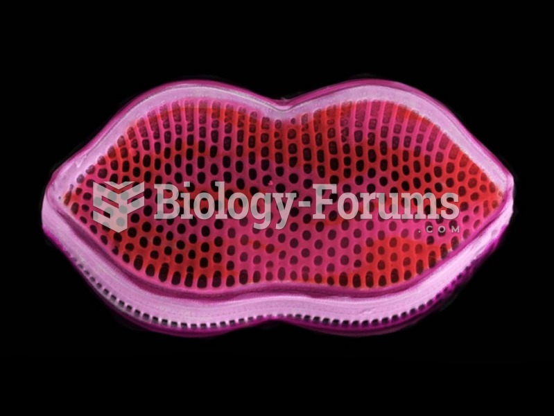 Electron Microscope Images of a Diatom
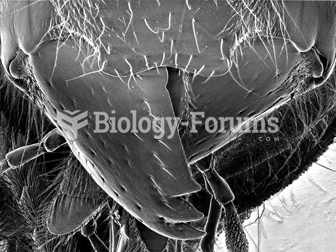 Scorpions, Spiders and Sharks: Electron-Microscope Images
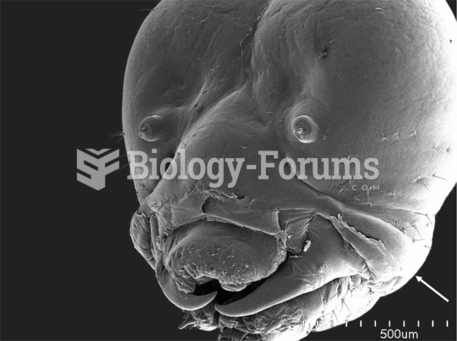 Scorpions, Spiders and Sharks: Electron-Microscope Images
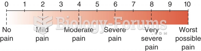 Numerical pain level chart with word modifiers.
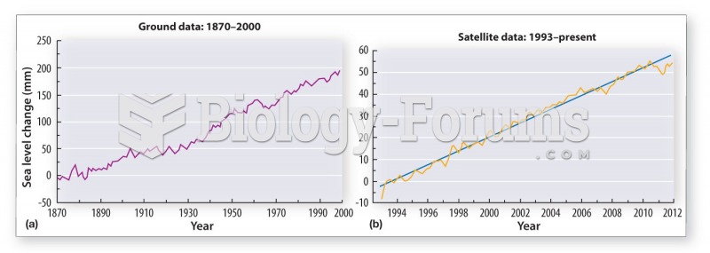 Rising sea level threatens coastal communities around the world
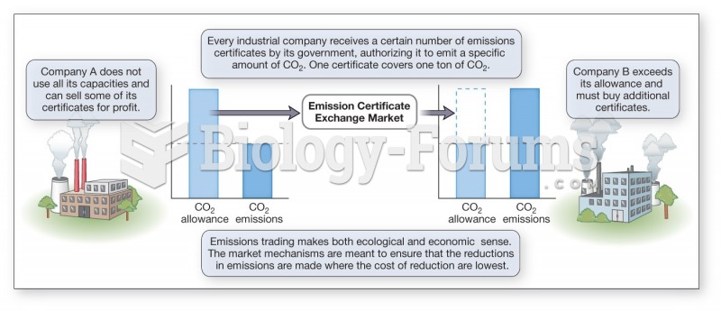 Kyoto Protocol encourages industry to innovate and reduce the level of emissions